JUNE 2, 1998 - THE HEART CATHETERIZATION
(And Unexpected Balloon Angioplasty)
Today was a big day. Ben had his first heart catheterization, or "cath", done. This procedure is used to provide detailed information about the condition of the heart. It shows how the heart chambers, valves and vessels are formed, and how they are functioning. This information is then used to determine if and when Ben is ready for his next operation, the Glenn.
We arrived at Children's Hospital at 6:30am. Ben was sedated and given a local anesthetic to numb the groin area. Once in the cath lab, a thin, flexible tube, known as a catheter, was inserted into an artery in his groin. Once the catheter was in the blood vessel, Dr. Singh, our WONDERFUL pediatric cardiologist, used a fluoroscope (similar to an x-ray machine) to guide the catheter into the different areas of Ben's heart.
While the catheter was in Ben's heart, several procedures were performed. Blood pressures in different heart chambers and blood vessels were recorded. Oxygen content of the blood in each heart chamber was evaluated. Dye was injected through the catheter. Angiograms (x-ray movies of the dye's movement) were filmed so that the details of any cardiac problem were recorded.
We were told it could take 1-4 hours. The cath did not start until 8:30, but much to our surprise (and joy), Dr. Singh was done by 10am.
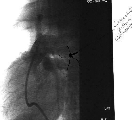
The results were mixed. The good news was that the pulmonary arteries looked great, which plays a big factor in the success of the Glenn. The bad news was that there was a newly discovered and unexpected problem called COARCTATION OF THE AORTA. This is a narrowing in the aorta which is restricting the blood flow out to the body. The majority of the aorta was about 8mm wide, but at the narrowing, it was just 2mm wide. You can see this in the photo below. (Since no dye had been injected during the cath yet, it was hard to see, so I outlined the problem area.)
|
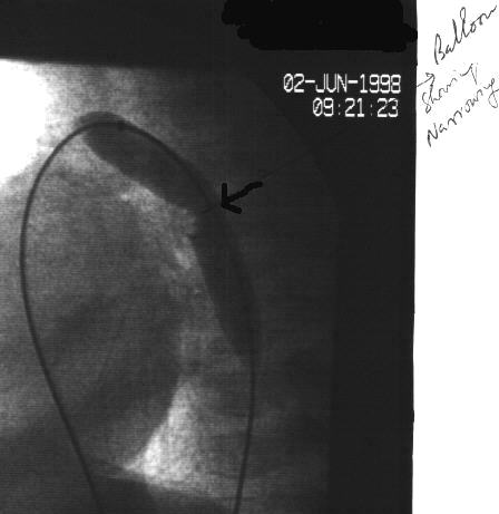
Dr. Singh decided to try to fix the coarctation using balloon angioplasty. The object of this is to blow up a balloon like instrument inside the walls of the aorta, in effect, stretching the inner layers and widening it.
The next picture shows the aorta with the balloon inside it, but not fully inflated. You can see the narrowing better in this picture.
|
The photo below shows the aorta with the balloon fully dilated or inflated. The object is to "tear" the inner layers of the aorta to widen it, without tearing the outer layers.
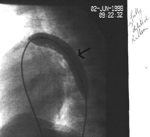 |
|
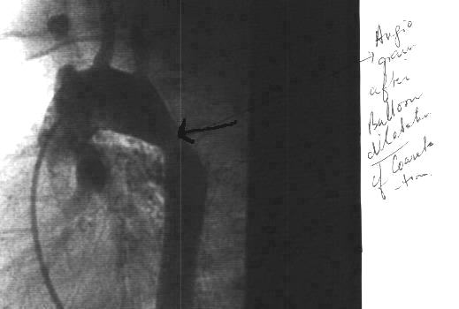
We hope this corrects the coarctation permanently. If it does not, Benjamin will require an additional surgery to correct this before he has his Glenn. Our next appointment is 6/9/98, so we will keep you up to date.
![]()